Wednesday, October 5, 2016

Chronic Wound Mycobiome

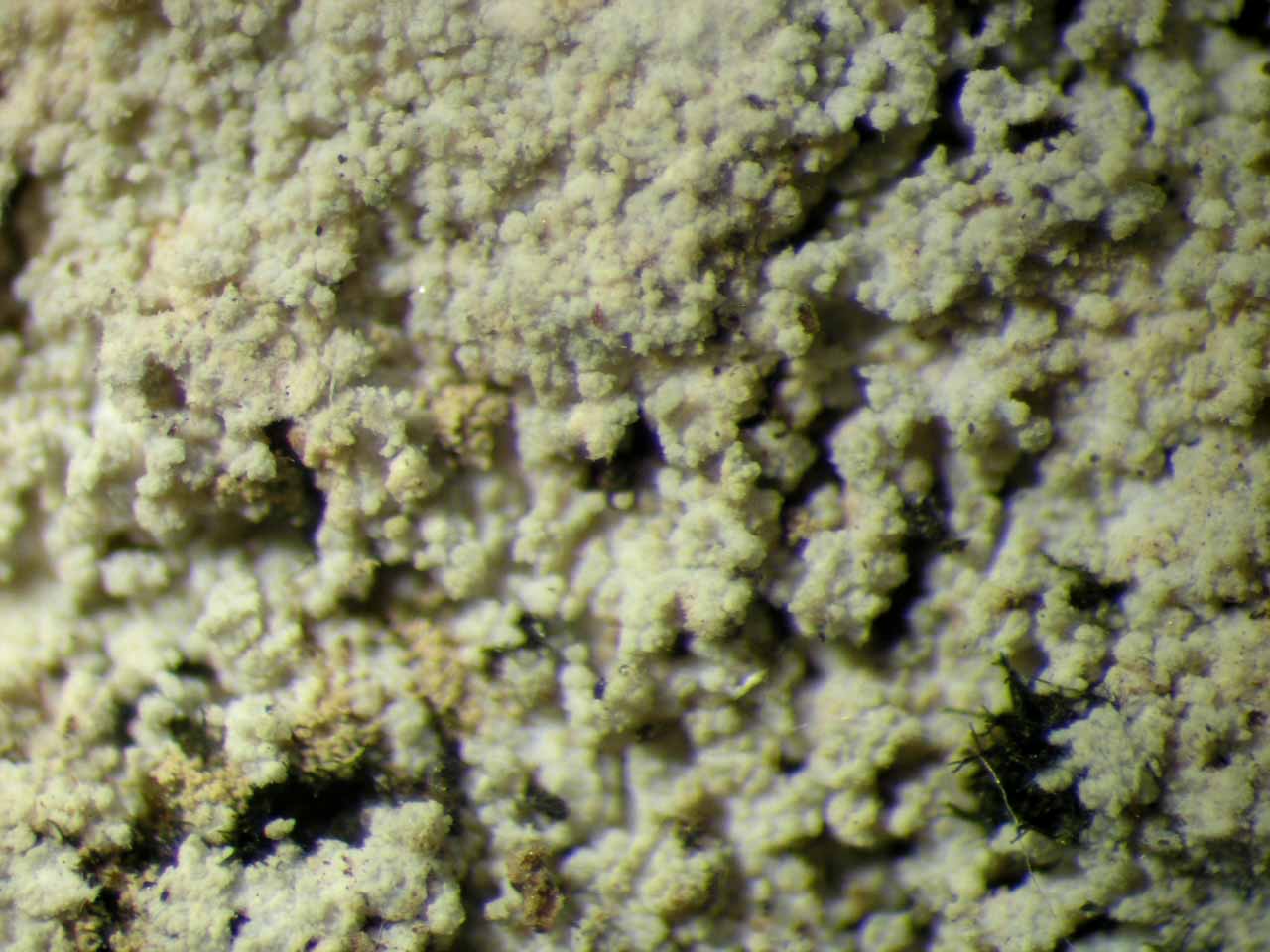
A new paper has just come out in mBio, and it represents an exploration of the fungi associated with chronic foot wounds in diabetic patients. This paper represents the last project for which I did benchwork before becoming all 'computational' all the time! I designed the approach for using MiSeq to sequence fungal ITS1 (which included designing and testing new primers specific for the platform) and executed the benchwork along with the first round of computational analyses. I appreciate Michael Loesche and Lindsay Kalan picking it up and performing a lot of necessary work to properly finish it off after I moved on from my position in the Grice Lab at the University of Pennsylvania.

The major finding was that certain aspects of the 'mycobiome' (i.e., the full set of fungi present) are associated with delayed healing, indicating that fungal profiling could become an important aspect of chronic wound care as medicine moves forward. Have a look at the link at the bottom of this post!

Pathogens are associated with poor outcomes. Mean proportion of pathogens (y axis) by end of study reason (x axis). Error bars indicate standard errors of the means.


--------------

Reference:

Kalan, L., M. A. Loesche, B. P. Hodkinson, K. P. Heilmann, G. Ruthel, S. E. Gardner, and E. A. Grice. 2016. Redefining the chronic wound microbiome: fungal communities are prevalent, dynamic, and associated with delayed healing. mBio 7(5): e01058-16.
Download publication (PDF file)

Friday, July 15, 2016

Cancer and HIV

We are at a point in time when the field of cancer research is becoming dominated by immunoncology - the study of how cancer interacts with the immune system.  One interesting aspect of this is that certain viral infections and certain cancer types might be most effectively treated using some of the same cellular and molecular machinery.  This CNN article addresses this in the context of HIV + acute myeloid leukemia and specifically mentions the generation and maintenance of CD8 T-cells as one way of combating both viral infections and specific types of cancer:
http://www.cnn.com/2016/07/15/health/cancer-research-hiv-cure/

- Brendan

Monday, February 29, 2016

Why Design Matters

Recently an article resulting from some of my research at the University of Pennsylvania was published.  It highlights how methods, approach, and experimental design can heavily influence the conclusions of studies on microbial communities.  Specifically, it shows that the most common primers used for microbiome studies (amplifying the 16S-V4 region) do a poor job of amplifying DNA from certain crucial microbes in the skin microbiome (such as Propionibacterium), biasing results and giving an overall inaccurate picture of skin microbial diversity.  A related observation was that the species of Staphylococcus could not be reliably identified using the V4 region, as shown here:

Relative abundance of Staphylococcus sequences able to be classified at the species level. (Top) Staphylococcus species in the Whole Metagenomic Sequence dataset were classified using MetaPhlAn. (Middle and Bottom) 16S-V1-V3 and 16S-V4 species level classifications were determined by pplacer. Pie charts depict the percentage of sequences classified as Staphylococcus at the genus level that were further classified at the species level.

To read a lengthier summary of the article, check out my friend and co-author Geof Hannigan's blog post!

- Brendan

---------------------------------------

Reference

Meisel, J., G. D. Hannigan, A. Tyldsley, A. J. SanMiguel, B. P. Hodkinson, Q. Zheng, and E. A. Grice. 2016. Skin microbiome surveys are strongly influenced by experimental design. Journal of Investigative Dermatology DOI: 10.1016/j.jid.2016.01.016.
Download manuscript (PDF file)

Tuesday, December 8, 2015

Citations

A quick look at Google Scholar the other day showed me that there have been over 2300 citations to my papers. The other citation metrics were interesting, too, like the h-index (currently 24, meaning that 24 of my papers have been cited 24 times or more) and the i10 index (currently 39, meaning that I have had 39 papers cited 10 times or more). These numbers only take into account the peer-reviewed publications, but I do have quite a number of additional non-peer-reviewed works. If I count up the peer-reviewed papers plus my other types of publications (abstracts, book reviews, annotated bibliographies, etc.), I now have well over 100 publications!

As an aside, one metric having to do with publication that is commonly used is the 'impact factor.' Although this number is meant to evaluate journals as a whole, it is often used by institutions to evaluate researchers, using the journals in which they publish as a proxy for their overall impact as scientists. This is, of course, highly controversial. One aspect that makes it even more problematic is that metrics like impact factor can be highly dependent on the database used for compiling the raw citation data. My wife published a paper a few years back in which this issue was explored quantitatively (Gray & Hodkinson 2008). In a set of 50 journals, many rankings were seen to differ by numbers that ran up into the double digits depending on the database used, calling into question the accuracy of any given journal's impact factor.

- Brendan

--------------------------

Reference

Gray, E, and S. Z. Hodkinson. 2008. Comparison of Journal Citation Reports and Scopus Impact Factors for Ecology and Environmental Sciences Journals. Issues in Science and Technology Librarianship DOI:10.5062/F4FF3Q9G.
View publication (website)

---------------------------

Note: Citation values updated May 29, 2018

Saturday, November 21, 2015

Skin Virome

The human skin is covered in many microbes, and until recently, the viral component has remained poorly understood. A recent paper presents the research into the human skin virome that I worked on while in the Dermatology Department at the University of Pennsylvania.

A cross-section of the skin (showing viruses and other microbes).

Thanks to Geof Hannigan for conceptualizing and taking the lead on this project. You can read his description of it and an overview of the results here:
http://prophage.blogspot.com/2015/10/new-study-looking-at-viruses-colonizing.html

There is also a good article for the general public here that describes the work:
http://arstechnica.com/science/2015/11/vast-uncharted-viral-world-discovered-on-human-skin/

- Brendan

-----------------

Reference

Hannigan, G. D., J. S. Meisel, A. S. Tyldsley, Q. Zheng, B. P. Hodkinson, A. J. SanMiguel, S. Minot, F. D. Bushman, and E. A. Grice. 2015. The human skin double-stranded DNA virome: topographical and temporal diversity, genetic enrichment, and dynamic associations with the host microbiome. mBio 6(5): e01578-15.
Download publication (PDF file)

Saturday, October 17, 2015

patPRO: Visualizing Longitudinal Microbiome Data

Recently some of my collaborators from the University of Pennsylvania and I released a new R package on CRAN (Comprehensive R Archive Network). It is called 'patPRO' (short for 'Patient Profiler'), and provides a number of functions to facilitate the visualization of longitudinal microbiome data. Although we developed it for examining changes in human microbiomes over time, it could just as easily be used for changes in microbial populations in water supplies, agricultural soils, or livestock, just to name a few possibilities.

This is an example of a plot that can be made, simultaneously showing changes in multiple dimensions of the microbiome:


Here is the official page for the package, with links to the reference manual and source code:
http://cran.us.r-project.org/web/packages/patPRO/index.html

This link provides a version of the reference manual in a series of webpages:
http://rpackages.ianhowson.com/cran/patPRO/

- Brendan

-------------------------------------

Reference

Hannigan, G. D., M. A. Loesche, B. P. Hodkinson, S. Mehta, and E. A. Grice. 2015. patPRO: Visualizing Temporal Microbiome Data. R package version 1.0.0. http://cran.r-project.org/web/packages/patPRO/index.html
Download package (website)

-------------------------------------

Note: One implementation of R gave me an error indicating that 'stringi' was needed when I tried to install 'patPRO.' After installing 'stringi,' I ran across no further issues with patPRO.

Saturday, September 26, 2015

Insights into Parmeliaceae

The results of a large-scale collaborative Parmeliaceae systematics project, in which I participated, were recently published in New Phytologist. To read the article, check out the link at the bottom of this post; I have pasted the abstract below.
  • We studied the evolutionary history of the Parmeliaceae (Lecanoromycetes, Ascomycota), one of the largest families of lichen-forming fungi with complex and variable morphologies, also including several lichenicolous fungi.
  • We assembled a six-locus data set including nuclear, mitochondrial and low-copy protein-coding genes from 293 operational taxonomic units (OTUs).
  • The lichenicolous lifestyle originated independently three times in lichenized ancestors within Parmeliaceae, and a new generic name is introduced for one of these fungi. In all cases, the independent origins occurred c. 24 million yr ago. Further, we show that the Paleocene, Eocene and Oligocene were key periods when diversification of major lineages within Parmeliaceae occurred, with subsequent radiations occurring primarily during the Oligocene and Miocene.
  • Our phylogenetic hypothesis supports the independent origin of lichenicolous fungi associated with climatic shifts at the Oligocene–Miocene boundary. Moreover, diversification bursts at different times may be crucial factors driving the diversification of Parmeliaceae. Additionally, our study provides novel insight into evolutionary relationships in this large and diverse family of lichen-forming ascomycetes.
- Brendan

-------------------------

Reference

Divakar, P. K., A. Crespo, M. Wedin, S. D. Leavitt, L. Myllys, B. McCune, T. Randlane, J. W. Bjerke, Y. Ohmura, I. Schmitt, C. G. Boluda, D. Alors, B. Roca-Valiente, R. Del-Prado, Constantino Ruibal, K. Buaruang, J. Núñez-Zapata, G. Amo de Paz, V. J. Rico, M. C. Molina, J. A. Elix, T. L. Esslinger, I. K. K. Tronstad, H. Lindgren, D. Ertz, C. Gueidan, L. Saag, T. Tõrra, G. Singh, F. Dal Grande, S. Parnmen, A. Beck, M. N. Benatti, D. Blanchon, M. Candan, P. Clerc, T. Goward, M. Grube, B. P. Hodkinson, J.-S. Hur, G. Kantvilas, P. M. Kirika, J. Lendemer, J.-E. Mattsson, M. I. Messuti, J. Miadlikowska, M. Nelsen, J. I. Ohlson, S. Pérez-Ortega, A. Saag, H. J. M. Sipman, M. Sohrabi, A. Thell, G. Thor, C. Truong, R. Yahr, D. K. Upreti, D. L. Hawksworth, P. Cubas, and H. T. Lumbsch. 2015. Evolution of complex symbiotic relationships in a morphologically derived family of lichen-forming fungi. New Phytologist DOI: 10.1111/nph.13553.
Download publication (PDF file)

Monday, August 31, 2015

New Job

I have officially started a new job at Janssen Research & Development, a member of the Johnson & Johnson family of companies! I am working within Oncology Translational Research as a Post-Doctoral Fellow in Computational Biology. With Janssen, I look forward to many opportunities to do groundbreaking research and really make an impact in people's lives!

- Brendan

Friday, July 31, 2015

PICS-Ord on Mac

In a previous post, I wrote about how to get the program PICS-Ord to run easily on a PC. Other operating systems presented some special problems associated with getting the different dependencies to find one another. I recently found Homebrew, which solves a lot of the problems typically found with Mac computers, and I put together some instructions for running PICS-Ord on the current Mac OS.

Running PICS-Ord on Mac OS X (10.9 or 10.10):

(1) Download and install R from here:
https://cran.r-project.org/bin/macosx/R-3.2.2.pkg
Open the file and follow the instructions.

(2) Install CMake and Boost using Homebrew; use the following three commands in the terminal to first install Homebrew and then the two Ngila dependencies:
ruby -e "$(curl -fsSL https://raw.githubusercontent.com/Homebrew/install/master/install)"
brew install cmake
brew install boost --with-python


(3) Install Ngila by using the following commands in the terminal (if you already have wget, you can ignore the first line):
brew install wget
wget http://scit.us/projects/files/ngila/Releases/ngila-release.tar.gz
tar zxvf ngila-release.tar.gz
cd ngila-1.3-release/
cmake . && make
make install
cd ..


(4) Download PICS-Ord from here:
http://scit.us/projects/files/ngila/picsord.zip
Open the zip file to create the picsord directory.

(5) Run PICS-Ord by moving the FASTA files into the picsord directory, navigating to the picsord directory in the terminal, and running the following:
Rscript picsord.R region_1.fas > region_1.phy
Rscript picsord.R region_2.fas > region_2.phy
Rscript picsord.R region_3.fas > region_3.phy
Rscript picsord.R region_4.fas > region_4.phy


- Brendan

-------------------------

Reference

Lücking, R., B. P. Hodkinson, A. Stamatakis, and R. A. Cartwright. 2011. PICS-Ord: Unlimited Coding of Ambiguous Regions by Pairwise Identity and Cost Scores Ordination. BMC Bioinformatics 12: 10.
Download publication (PDF file)
Download R-based program (zipped program package)
View program wiki (website)

Friday, June 12, 2015

Shared Microbiota of Humans and Their Pets

I recently co-authored a paper from a study in which we examined the microbiota of humans and their pets.  The full text can be seen here:

Abstract -

Background: Staphylococcus aureus and other coagulase-positive staphylococci (CPS) colonize skin and mucous membrane sites and can cause skin and soft tissue infections (SSTIs) in humans and animals. Factors modulating methicillin-resistant S. aureus (MRSA) colonization and infection in humans remain unclear, including the role of the greater microbial community and environmental factors such as contact with companion animals. In the context of a parent study evaluating the households of outpatients with community MRSA SSTI, the objectives of this study were 1) to characterize the microbiota that colonizes typical coagulase-positive Staphylococcus spp. carriage sites in humans and their companion pets, 2) to analyze associations between Staphylococcus infection and carriage and the composition and diversity of microbial communities, and 3) to analyze factors that influence sharing of microbiota between pets and humans.

Results:We enrolled 25 households containing 56 pets and 30 humans. Sampling locations were matched to anatomical sites cultured by the parent study for MRSA and other CPS. Bacterial microbiota were characterized by sequencing of 16S ribosomal RNA genes. Household membership was strongly associated with microbial communities, in both humans and pets. Pets were colonized with a greater relative abundance of Proteobacteria, whereas people were colonized with greater relative abundances of Firmicutes and Actinobacteria. We did not detect differences in microbiota associated with MRSA SSTI, or carriage of MRSA, S. aureus or CPS. Humans in households without pets were more similar to each other than humans in pet-owning households, suggesting that companion animals may play a role in microbial transfer. We examined changes in microbiota over a 3-month time period and found that pet staphylococcal carriage sites were more stable than human carriage sites.

Conclusions: We characterized and identified patterns of microbiota sharing and stability between humans and companion animals. While we did not detect associations with MRSA SSTI, or carriage of MRSA, S. aureus or CPS in this small sample size, larger studies are warranted to fully explore how microbial communities may be associated with and contribute to MRSA and/or CPS colonization, infection, and recurrence.

--------------------------------------------

Reference

Misic, A. M., M. F. Davis, A. S. Tyldsley, B. P. Hodkinson, P. Tolomeo, B. Hu, I. Nachamkin, E. Lautenbach, D. O. Morris, and E. A. Grice. 2015. The shared microbiota of humans and companion animals as evaluated from Staphylococcus carriage sites. Microbiome 3: 2.
Download publication (PDF file)

Saturday, November 1, 2014

Next-Gen Sequencing: A Review

I recently wrote up a review with Dr. Elizabeth Grice of next-generation sequencing technologies. We specifically focused on their applications for microbiome research. While it was published it in a wound care journal, the techniques outlined are broadly applicable to any system with a diversity of microbes. Please have a look at the manuscript below!

-----------

Reference

Hodkinson, B. P., and E. A. Grice. 2015. Next-generation sequencing: a review of technologies and tools for wound microbiome research. Advances in Wound Care 4(1): 50-58.
Download manuscript (PDF file)

Saturday, October 25, 2014

Mole Day

This year I am teaching several sections of High School Chemistry, and this past week we had celebrations for Mole Day!  Here's a rundown of some of the snacks and activities.

Moles May Attend:
Make A Mole - For extra credit students can make stuffed animal moles from a pattern (just Google it). It's great to have some homemade moles in attendance at the party!

Food:
Guacamole - Trader Joe's has a guacamole dip called "Avocado's Number" that actually has a picture of Amadeo Avogadro on it, which is a great prop. I also decided to make fresh guacamole for the students, which provided everyone with a bit of a show.
Molasses cookies - OK, so the only real connection here is that 'mol' is in the word, but who doesn't like cookies?!

Sing-Along:
Rock Me Amadeo - The song 'Rock Me Amadeus' by Falco has an awesome video that highlights the time-period in Europe when Amadeo Avogadro was alive (which overlaps with the time period when Wolfgang Amadeus Mozart was alive, which is the reason that the video is set in this time period). Students can sing 'Amadeo' instead of 'Amadeus' on the chorus and you've got a catchy theme song for Mole Day!

Game Show:
More Than, Less Than, or Equal to a Mole - You can take objects that are basically made of one type of compound and ask students if they are more than a mole, less than a mole, or equal to a mole. Students can be split into two teams, and pairs (with one from each team) can stand on either side of a bell and compete to ring in first. If the first to answer is incorrect, the other person gets a chance to guess. I have to give credit to Mrs. Swieson (also of Delaware County Christian School) for coming up with this one!

Most of all, we just partied and had a great time hanging out as a class!

Saturday, September 6, 2014

Electronic Publication of Taxa


Hodkinson, B. P., and J. C. Lendemer. 2014. A clarification of effective electronic publication. Taxon 63(4): 911-913.
Download publication (PDF file)

Saturday, August 23, 2014

Lecanoromycetes

Please check out the newest publication that we've put together on the phylogeny and taxonomy of the fungal class Lecanoromycetes (the main group of lichen-forming fungi). This publication is more all-encompassing than any before it, including analyses of molecular sequence data from 1307 different fungi!

Miadlikowska, J., F. Kauff, F. Högnabba, J. C. Oliver, K. Molnár, E. Fraker, E. Gaya, J. Hafellner, V. Hofstetter, C. Gueidan, M. A. G. Otálora, B. Hodkinson, M. Kukwa, R. Lücking, C. Björk, H. J. M. Sipman, A. R. Burgaz, A. Thell, A. Passo, L. Myllys, T. Goward, S. Fernández-Brime, G. Hestmark, J. Lendemer, H. T. Lumbsch, M. Schmull, C. L. Schoch, E. Sérusiaux, D. R. Maddison, A. E. Arnold, S. Stenroos, and F. Lutzoni. 2014. A multigene phylogenetic synthesis for the class Lecanoromycetes (Ascomycota): 1307 fungi representing 1139 infrageneric taxa, 317 genera and 66 families. Molecular Phylogenetics and Evolution 79: 132-168.
Download publication (PDF file)
Download supplementary file 1 (PDF file)
Download supplementary file 2 (PDF file)
Download supplementary file 3 (PDF file)
Download supplementary file 4 (PDF file)
Download supplementary file 5 (docx file)
Download supplementary file 6 (PDF file)
Download supplementary file 7 (docx file)

Friday, July 11, 2014

Spider Bite


[WARNING: Graphic Content]

Over the past month, my wife has been dealing with an ulcer on her shoulder that was most likely caused by a spider bite (I'm guessing that some type of Sac Spider is to blame). At first, it raised up like a volcano with a base diameter of about 2-3 cm and a height of about 1-2 cm. Then after a couple of days, the center became necrotic (see "June 15" below). She had the part that was entirely necrotic taken out by a doctor, leaving a bit of a hole in the center ("June 17"). She then treated it daily with honey and kept it covered for the next few weeks. During this time, there was a period of a few days where the whole area started to become red and inflamed ("July 1"), so she got on an antifungal (Fluconazole) and two antibiotics (Keflex & Minocycline), which quickly cleared up that particular issue. It is now healing up nicely ("July 4") and we hope it will finish up without any additional complications!


Timeline (all treatments follow doctors' recommendations):
June 12 - noticed painful swelling
June 15 - Keflex treatment begun (for potential general infection)
June 16 - Keflex treatment terminated, Bactrim treatment begun (for potential MRSA infection)
June 17 - necrotic center removed/biopsied, treatment with honey begun
June 19 - Bactrim treatment terminated (no signs of infection from lab tests)
July 1 - Keflex, Minocycline and Fluconozole treatment begun (swelling and redness seen around the area)


Monday, July 7, 2014

Sticta sylvatica

I recently had a paper published that talks about the status of the rare species Sticta sylvatica in my part of the world (Hodkinson et al. 2014). The article was actually featured on the cover! Here's the abstract:

"The presence of the foliose cyanolichen Sticta sylvatica in eastern North America has been called into question due to the absence of high-quality, verifiable material and the common misuse of its name. Recently, specimens collected in the Great Smoky Mountains have been verified as having the typical S. sylvatica morphology. Although molecular data remain inconclusive regarding the entity’s genetic distinctiveness from the phenotypically dissimilar S. limbata, we argue that the decline in the abundance of this morphological entity worldwide along with the need for further genetic study make continued conservation efforts imperative."

Sticta sylvatica in the field.

- Brendan

---------------------

Reference

Hodkinson, B. P., J. C. Lendemer, T. McDonald, and R. C. Harris. 2014. The status of Sticta sylvatica, an ‘exceedingly rare’ lichen species, in eastern North America. Evansia 31(1): 17-24.
Download publication (PDF file)
Download journal issue cover (PDF file)

---------------------

[This work was supported by the National Science Foundation under awards EF-1115086 and DEB-1145511.]


Monday, June 30, 2014

Molecular Ecology

A few years back, I began working on a collaborative project led by Greg Bonito to examine fungal and bacterial communities associated with plant roots.  We used next-generation sequencing on the 454 GS-FLX platform for profiling using four different loci (one bacterial and three fungal).  The fungal ITS and LSU primers were identical to those used in my recent Mycosphere paper (Hodkinson & Lendemer 2013), and the 16S primers are the ones I designed for my Environmental Microbiology paper from a couple of years back (Hodkinson et al. 2012).  The paper is finally out in Molecular Ecology; check it out!

- Brendan

------------------------

References

Bonito, G., H. Reynolds, M. S. Robeson, J. Nelson, B. P. Hodkinson, G. Tuskan, C. W. Schadt, and R. Vilgalys. 2014. Plant host and soil origin influence fungal and bacterial assemblages in the roots of woody plants. Molecular Ecology 23(13): 3356-3370.
Download publication (PDF file)

Hodkinson, B. P., and J. C. Lendemer. 2013. Next-generation sequencing reveals sterile crustose lichen phylogeny. Mycosphere 4(6): 1028-1039.
Download publication (PDF file)
Download data and sequence-processing scripts (ZIP archive)
Download Ascomycota LSU alignment and analysis files (ZIP archive)
Download Arthoniales LSU alignment and analysis files (ZIP archive)

Hodkinson, B. P., N. R. Gottel, C. W. Schadt, and F. Lutzoni. 2012. Photoautotrophic symbiont and geography are major factors affecting highly structured and diverse bacterial communities in the lichen microbiome. Environmental Microbiology 14(1): 147-161.
Download publication (PDF file)
View publication (publisher's website)Download supplementary phylogeny (PDF file)
Download data and analysis file archive (ZIP file)

------------------------

[Support for my work on this project was provided in part by the National Science Foundation under awards EF-0832858, DEB-1011504, and DEB-1145511.]

Wednesday, April 30, 2014

Dynamic Periodic Table

Here's a really cool dynamic periodic table that nicely lays out all sorts of information about the various elements (with all sorts of tabs and sliders):
I wish there had been something like this when I was in high school and college!

Wednesday, March 26, 2014

Open Fracture Microbiome

I recently co-authored a paper with a team of dermatologists (those who study skin) and orthopaedists (those who study bone) in which we present the results of microbiome analyses on open fracture wounds. These are the types of wounds where bones actually break through the surface of the skin. We found that the wound-associated microbial communities change over time, becoming significantly more similar to the adjacent skin communities as healing progresses. There are also a number of bacterial groups and aspects of the microbiome that tend to be associated different clinical factors. Please have a look at the publication at the links below!

- Brendan

----------------------

Reference

Hannigan, G. D., B. P. Hodkinson, K. McGinnis, A. S. Tyldsley, J. B. Anari, A. D. Horan, E. A. Grice, and S. Mehta. 2014. Culture-independent pilot study of microbiota colonizing open fractures and association with severity, mechanism, location, and complication from presentation to early outpatient follow-up. Journal of Orthopaedic Research 32(4): 597-605.
Download publication (PDF file)
Download supplemental methods (PDF file)
Download sample metadata (QIIME mapping file)
Download OTU table (bzip2-compressed BIOM file)
Download OTU representative sequences (bzip2-compressed FASTA file)

----------------------

[This work was funded in part by NIAMS/NIH R00 AR060873.]

Friday, February 28, 2014

Alternative Nitrogen Fixation

During my dissertation research, I found an interesting set of genes in the metatranscriptome of Peltigera praetextata. They were vnfDG and vnfN, two genes in the alternative, vanadium-dependent nitrogen fixation gene cluster. The interesting thing about this was that nitrogen fixation in lichens has always been attributed to the standard molybdenum-dependent system. These gene fragments were very similar to ones from Anabaena, cyanobacteria that are not known to be lichen photobionts, but are closely related to Nostoc, the cyanobiont in Peltigera. After examining metagenomes from other Peltigera species (all associated with Nostoc) and running further analyses, I found that the presence of the vnf gene cluster seems to be widespread (in all five samples checked from five Peltigera species from all over the world). Analyses revealed that the Peltigera-associated sequences all form a group close to, but separate from, Anabaena, which is entirely consistent with them being derived from the main photobiont, Nostoc. Therefore, it seems that lichens with cyanobacteria are actually using both the standard nitrogen-fixation system and an alternative, vanadium-dependent one.

This work could have sweeping implications for studies of biogeochemistry, since certain ecosystems, especially those in the tundra, are dominated by lichens. Ecosystem-wide nitrogen fixation rates are often inferred based on the acetylene-reduction assay (ARA). To interpret the results of this assay, a conversion factor based on the standard molybdenum-dependent system is typically used. However, if a substantial portion of the nitrogen fixation is taking place via alternative means, the standard conversion factor could produce wildly inaccurate interpretations. Even if one is willing to consider that a different conversion factor should be used, determining the proper conversion factor may become especially problematic since the proportion of standard to alternative nitrogen fixation could vary greatly based on the season or micro-environment. Therefore, a greater understanding of vanadium, molybdenum, and nitrogen dynamics may be needed before we can continue to rely blindly upon the commonly-used ARA for ecosystem-wide studies.

Peltigera rufescens, a lichen with a cyanobacterial photobiont from the genus Nostoc, in the Alaskan tundra.

- Brendan

------------------------

Reference

Hodkinson, B. P., J. L. Allen, L. L. Forrest, B. Goffinet, E. Sérusiaux, Ó. S. Andrésson, V. Miao, J.-P. Bellenger, and F. Lutzoni. 2014. Lichen-symbiotic cyanobacteria associated with Peltigera have an alternative vanadium-dependent nitrogen fixation system. European Journal of Phycology 49(1): 11-19.
Download publication (PDF file)
Download supplementary table (PDF file)
Download vnfD alignment and analysis files (ZIP archive)
Download vnfN alignment and analysis files (ZIP archive)
Download script for editing GenBank-derived FASTA files (PERL script)

------------------------

[This work was supported in part by the National Science Foundation under grants OCI-1053575 and DEB-0919284.]

Saturday, February 15, 2014

New Species: Lepidostroma winklerianum

There has recently been amazing burst of discovery in what is now known as the order Lepidostromatales. Until 2007, only one of the species that is now in the order had been described. Then, in a series of recent papers, five new species have been described. The most recent new species is Lepidostroma winklerianum, named after Sieghard Winkler, one of the authors who first described the genus Lepidostroma.

Morphology and anatomy of thallus squamules of Lepidostroma winklerianum
(a–b) Fully grown and young squamules. [scale=1 mm] 
(c) Section through squamule.  [scale=100 μm]
(d) Photobiont cells.  [scale=10 μm]
(e) Upper cortex in section view. [scale=10 μm]
(f) Upper cortex in surface view. [scale=10 μm]

Here is a key to the currently-known species in the order Lepidostromatales:

1a Thallus crustose, undifferentiated, lacking distinct cortical structures (Sulzbacheromyces); basidiomata clavarioid; Neotropics (northeastern Brazil) ... Sulzbacheromyces caatingae
1b Thallus microsquamulose to squamulose, with distinct cellular cortex; basidiomata clavarioid or club-shaped ... 2

2a Thallus microsquamulose, composed of contiguous glomerules with cortex formed by distinctly lobate, jigsaw-puzzle-shaped cells (Ertzia), medulla absent; basidiomata clavarioid; tropical Africa (Rwanda) ... Ertzia akagerae
2b Thallus distinctly squamulose, composed of scattered to dense, rounded to reniform squamules with cortex formed by polygonal or jigsaw-puzzle-shaped cells (Lepidostroma), medulla present; basidiomata clavarioid or club-shaped ... 3

3a Basidiomata clavarioid; squamules lacking raised margin and maculae; photobiont layer above medulla, more or less uniform, with scattered cells throughout medulla and in lower portion of squamules; photobiont cells lacking pyrenoids; Neotropics (Costa Rica, Colombia) ... Lepidostroma calocerum
3b Basidiomata club-shaped; squamules with conspicuous, slightly raised white margin, their surface maculate; photobiont layer below medulla, forming pyramidal columns protruding upwards; photobiont cells with pyrenoid(s) ... 4

4a Upper cortex with jigsaw-puzzle-shaped cells in surface view; tropical Africa (Rwanda) ... Lepidostroma rugaramae
4b Upper cortex with polygonal cells in surface view; Neotropics (Mexico) ... 5

5a Squamules 1.5–3 mm diam.; upper cortex multi-layered; basidia 2-sterigmate ... Lepidostroma vilgalysii
5b Squamules 0.5–1.5(–2) mm diam.; upper cortex single-layered; basidia 4-sterigmate ... Lepidostroma winklerianum

Brendan

-------------
Hodkinson, B. P., B. Moncada, and R. Lücking. 2014. Lepidostromatales, a new order of lichenized fungi (Basidiomycota, Agaricomycetes), with two new genera, Ertzia and Sulzbacheromyces, and one new species, Lepidostroma winklerianumFungal Diversity 64(1): 165-179.
Download publication (PDF file)
View publication (website)
-------------

[This work was supported in part by the National Science Foundation under DEB-0715660.]

Friday, January 24, 2014

New Genera: Ertzia & Sulzbacheromyces

For a recent paper establishing the new order Lepidostromatales, we examined the morphological and molecular diversity of the genus Lepidostroma, and it became clear that there were three very different types of species. The core group of four species has rounded to reniform squamules. However, one species is outside of this group and has a microsquamulose thallus that forms contiguous glomerules with a cortex of jig-saw-puzzle-shaped cells. We gave this species its own genus and called it Ertzia akagerae, naming the genus after Damien Ertz, the primary describer of the species. Another species outside of the core group has an entirely crustose thallus, so we gave it a new genus and named it Sulzbacheromyces caatingae. The genus was named for Marcelo Sulzbacher who described the single species with colleagues. Although it's still inconclusive, there also could be photobiont differences associated with the split of these three genera. The samples of Lepidostroma and Sulzbacheromyces from which we were able to obtain algal DNA reads yielded sequences that were very different from one another, but were both from groups not known to be associated with any other types of lichenized fungi.

Brendan

-------------
Hodkinson, B. P., B. Moncada, and R. Lücking. 2014. Lepidostromatales, a new order of lichenized fungi (Basidiomycota, Agaricomycetes), with two new genera, Ertzia and Sulzbacheromyces, and one new species, Lepidostroma winklerianumFungal Diversity 64(1): 165-179.
Download publication (PDF file)
View publication (website)
-------------

[This work was supported in part by the National Science Foundation under DEB-0715660.]

Tuesday, January 14, 2014

New Order: Lepidostromatales

Some colleagues from the Field Museum and I recently described a new order of fungi, Lepidostromatales (Hodkinson et al. 2014). One thing that makes it unique is that it is the only order of basidiomycete fungi containing only lichenized members. The ordinal placement of the family Lepidostromataceae had previously been uncertain, but our molecular analyses confirmed its isolated position and made it clear that the best treatment would be to give it its own separate order. Please have a look at the new paper in Fungal Diversity for a key to all of the species in the new order!

- Brendan

[Update: I decided to also post the key to the species here.]

-------------
Hodkinson, B. P., B. Moncada, and R. Lücking. 2014. Lepidostromatales, a new order of lichenized fungi (Basidiomycota, Agaricomycetes), with two new genera, Ertzia and Sulzbacheromyces, and one new species, Lepidostroma winklerianum. Fungal Diversity 64(1): 165-179.
Download publication (PDF file)
View publication (website)
-------------

[This work was supported in part by the National Science Foundation under DEB-0715660.]

Wednesday, December 11, 2013

Next-generation sequencing reveals sterile crustose lichen phylogeny

I recently had a paper published that showed the utility of next-generation sequencing and a microbiome-based approach for basic lichen-forming fungal systematics. DNA sequences derived from the species known as Lepraria moroziana had for many years either been too messy to read or had seemingly been derived from disparate fungi, presumably because the species contains many endo-lichenic associates and/or it often grows in close proximity to other lichen-forming fungi. However, when using 454 sequencing to examine the whole pool of LSU (nuclear ribosomal large sub-unit) sequences from ten samples, it became clear that the dominant fungus in each was from the class Arthoniomycetes. This provided a breakthrough for the group, which was previously classified in the class Lecanoromycetes. After further investigations, it became clear that the two species Lepraria moroziana and Lepraria obtusatica belonged in their own genus, Andreiomyces, which itself we put in its own family, Andreiomycetaceae.

Relative abundances of the various putative classes represented by LSU sequences in each of the ten samples from the species of interest.

Interestingly, there were some sequences found in some of the amplicon pools derived from the "real" genus Lepraria (Lepraria s. str. in the class Lecanoromycetes). Had we gotten these sequences through standard Sanger sequencing, we may have been led astray, making it more difficult to discern the fact that the main lichen-forming fungus belongs to a previously unknown lineage in the class Arthoniomycetes!

I anticipate that this type of methodology will be advantageous for a lot of problematic fungi, especially those that remain uncultured. Here is the full abstract for the associated paper:

"The rapid phylogenetic placement and molecular barcoding of fungi is often hampered in organisms that cannot easily be grown in axenic culture or manually teased apart from their associated microbial communities. A high-throughput procedure is outlined here for this purpose, and its effectiveness is demonstrated on a representative species from an especially problematic group of fungi, the sterile crustose lichens. Sequence data of the LSU and ITS regions were generated from samples of a sterile crustose lichen species, Lepraria moroziana, using next-generation sequencing. DNA fragments most likely to represent the primary lichen-forming fungus were bioinformatically teased out using a specialized data processing pipeline. Phylogenetic analyses of the LSU region revealed that the lichen-forming fungus L. moroziana was previously placed in the incorrect class of fungi (Lecanoromycetes), and actually belongs to the class Arthoniomycetes, in the order Arthoniales. It is here treated as a member of a new family (Andreiomycetaceae Hodkinson & Lendemer fam. nov.) and genus (Andreiomyces Hodkinson & Lendemer gen. nov.). Additionally, Lepraria obtusatica Tønsberg is placed in the newly-defined genus based on its morphological, chemical, and ITS-based molecular similarity to L. moroziana. The procedure outlined here is projected to be especially useful for resolving the dispositions of diverse problematic fungi that remain unnamed, incertae sedis, or have taxonomic positions that are not expected to reflect their true phylogeny."

- Brendan

-------------------------

Reference

Hodkinson, B. P., and J. C. Lendemer. 2013. Next-generation sequencing reveals sterile crustose lichen phylogeny. Mycosphere 4(6): 1028-1039.
Download publication (PDF file)
Download data and sequence-processing scripts (ZIP archive)
Download Ascomycota LSU alignment and analysis files (ZIP archive)
Download Arthoniales LSU alignment and analysis files (ZIP archive)

Friday, December 6, 2013

Leprocaulales

This year I published a paper with James Lendemer (Lendemer & Hodkinson 2013) in which we established a new order of fungi, Leprocaulales, for a group of sterile crustose lichens.  We first found that the species in the genera Lepraria and Leprocaulon were all shuffled up (they got this way due to their similar appearance), but we were able to sort them properly using molecular data.  It then became apparent, after further analyses, that the Leprocaulon clade is actually quite distantly related to other known groups of fungi (and is not even close to Lepraria), so we gave it a new family (Leprocaulaceae) and new order (Leprocaulales).  One really interesting aspect of this work is that it revealed to us that both groups (Lepraria and Leprocaulon) had crustose and fruticose growth forms within them.  This shows that growth form is more plastic than most of us had probably suspected, and that an entirely new growth form can evolve in fungi on a very short time scale!

Figure 1 from Lendemer & Hodkinson (2013): This tree shows the placement of Lepraria s.l. species (including the species of Leprocaulon) in multiple groups within four families of Ascomycetes. Newly generated sequences of Lepraria s.l. are mapped to the topology of the Schmull et al. (2011) Lecanoromycetidae phylogeny. All taxa with a "leprarioid" growth form are in red.

- Brendan

---------------------

Reference:

Lendemer, J. C., and B. P. Hodkinson. 2013. A radical shift in the taxonomy of Lepraria s.l.: molecular and morphological studies shed new light on the evolution of asexuality and lichen growth form diversification. Mycologia 105: 994-1018.
Download publication (PDF file)
View data and analysis file web-portal (website)

Friday, November 8, 2013

Science Lichen Spoof

Recently Science magazine spoofed a bunch of other journals to test their peer-review procedures. It revealed what one might call a very porous filtration system on the part of many journals. A fake study with some pretty obvious problems was accepted for publication in quite a number of journals. You can read more about it here. One thing that caught my attention was that the fake article was on lichens and their potential anti-cancer properties. It is worth noting that there have been several recent articles that were not part of the spoof that show some anti-cancer properties of Usnic Acid; however, it's always important to carefully evaluate the legitimacy of any particular study and remain skeptical until results have been widely validated!

Saturday, October 19, 2013

Lichen-Inspired Technology

This article shows how lichens are inspiring new technological advancements:
http://www.ncbi.nlm.nih.gov/pubmed/23881167

Here is one excerpt from the article:

"Inspired by crustose lichens, one of the most common lichens that can grow within rocks, with a root-like fungal structure growing within the rock itself and only a crust-like fruiting body above the surface, we designed a novel integrated counter electrode architecture, in which a porous carbon plate (PCP) was used as a conductive substrate and continuous, vertically oriented ordered mesoporous carbon (OMC) monolithic films were rooted in the porous substrate to serve as catalytic layers (Scheme 1). Morphologically, the main body of crustose lichens (thallus) is made up of a few distinct layers. The lowermost layer consists of densely packed fungal hyphae with a root-like structure (rhizines) that can attach the thallus to various substrates such as rocks, plants, buildings, and even coral reefs. The root-like structure not only enables the lichen to adhere strongly to the substrate, thus preventing it from being peeled off by the wind or waves, but it also ensures the symbiont a much larger active area for mineral and water uptake, and photosynthesis. Herein, instead of a condensed flat conductive substrate such as FTO or metal foil, a porous carbon plate with both high conductivity and high mechanical strength was used as the CE substrate. A root-like integrated OMC–C composite structure formed by a precursor inflation pyrolysis process can harvest the electrons that arrive at the counter electrode and transfer them to the catalytic layer/electrolyte interface efficiently."

Pretty cool stuff!

-Brendan

Tuesday, October 1, 2013

Copper-Infused Socks for Diabetic Foot Care

I just came across an article about a new type of therapy for diabetic foot ulcers.  It caught my attention especially because it is aimed at treating infections that are fungal in nature.  In short, the product is a new type of sock infused with copper, which is shown to help stop the growth of certain problematic fungi.  This is extremely relevant to the ongoing diabetic foot ulcer microbiome work that we're doing in the Grice Lab.  Hopefully this product will provide some relief and reduce complications for many diabetes patients suffering from infections that may be primarily fungal!

- Brendan

Monday, September 23, 2013

Against the naming of fungi

Here's an interesting opinion piece published in Fungal Biology entitled "Against the naming of fungi:"
http://www.sciencedirect.com/science/article/pii/S1878614613000871
It highlights problems with the Linnean taxonomic system, and proposes using a system of molecularly-based operational taxonomic units (OTUs) that are named in a standardized way.  While this is an interesting and appealing idea, it comes with its own problems.  I think that it is important for everyone to recognize the problems with the current system, but I cannot say I'm on board with any particular alternative.  We'll see how much traction this gets in years to come!

-Brendan

Monday, August 26, 2013

Nelsenium

In a recent paper on the genus Lepraria (Lendemer & Hodkinson 2013), my co-author and I established a new genus named 'Nelsenium' for Lepraria usnica, a corticolous, Paleotropical species (Sipman 2003) that had previously been misclassified. The name honors Matt Nelsen, a fellow North American lichenologist who was the first to sequence Lepraria usnica and conducted molecular phylogenetic analyses to determine its familial affinities within the Lecanoromycetes. When Nelsen et al. (2008) used mtSSU sequence data to infer the higher level systematic placement of Lepraria usnica, they recovered the species as a member of the Pilocarpaceae (which is not the family of Lepraria). In our recent paper we affirmed this based on an analysis of 11 mtSSU sequences of Pilocarpaceae including L. usnica. Nelsen et al., however, did not formally describe a new genus to accommodate this species, so we established the genus Nelsenium to place L. usnica in its proper context.

We used a molecular diagnosis for this genus, naming the particular nucleotide positions that were unique in the mtSSU region.
"Diagnosis: A genus of the Pilocarpaceae, with a sterile, leprose crustose thallus whose mtSSU sequence (AY300894) differs from those of Fellhanera bouteillei (AY567787) and F. subtilis (AY567786) at these nucleotide sites: adenine at positions 96, 106, 232, 355, 689, 760, 806 and 813; cystine at positions 146 and 741; guanine at positions 44, 101 and 292; and thymine at positions 83, 233, 254 and 909."
I think this type of approach will become more and more common, especially in fungal taxonomy, where synapomorphies are often very difficult to find (especially for higher-level taxa) if one does not resort to examining the primary structure of nucleic acids or proteins.

To see how the new genus looks, feel free to peruse these photos of the type specimen of the type species, Nelsenium usnicum (Sipman) Lendemer & Hodkinson:
http://www.bgbm.org/digitalimages/Singa/k-pictures/P1010776k.jpg
http://www.bgbm.org/digitalimages/Singa/k-pictures/P1010777k.jpg

- Brendan

---------------------
References

Lendemer, J.C., and B.P. Hodkinson. 2013. A radical shift in the taxonomy of Lepraria s.l.: molecular and morphological studies shed new light on the evolution of asexuality and lichen growth form diversification. Mycologia 105: 994-1018.
Download publication (PDF file)

Nelsen, M.P., H.T. Lumbsch, R. Lucking, and J.A. Elix. 2008. Further evidence for the polyphyly of Lepraria (Lecanorales: Stereocaulaceae). Nova Hedwigia 87: 361-371.

Sipman, H.J.M. 2003. New species of CryptotheciaLepraria, and Ocellularia (lichenized Ascomycetes) from Singapore. Bibliotheca Lichenologica 86: 177-184.

Monday, August 19, 2013

Katydid Lichen Mimic

Here's a cool new species of katydid that looks like a lichen!
http://www.isidewith.com/article/new-to-nature-no-112-lichenagraecia-cataphracta

-Brendan

Sunday, July 28, 2013

Beatrix Potter

Today is the 147th birthday of Beatrix Potter.  Although she is best know for her books geared toward children, she was also an amateur mycologist/lichenologist and produced many drawings of fungi.  Here is one lichen drawing that she did.  She was also a big proponent of the dual nature of lichens (i.e., that they represent a symbiosis of fungi and algae), which was still controversial in her day.  You can read more about her scientific pursuits here!

-Brendan

Thursday, June 27, 2013

Lichens: A Cure for Cancer?

As the current overseer of recent lichen literature, I have noticed a number of articles that have come out in the past year or so demonstrating the anticancer effects of lichen compounds, specifically usnic acid.

Here are a few recent titles with links to articles:

It will be fascinating to see where this might lead!

Friday, June 21, 2013

mothur MiSeq SOP

As someone who uses the MiSeq platform for high-throughput DNA amplicon sequencing, I was excited to see a new paper in Applied & Environmental Microbiology that presents a pipeline for processing these data. Users of the program "mothur" were sent an email announcing the publication:

"Hello mothur users...
I know many of you have been interested in our protocols and the ideas behind our new MiSeq SOP. We have some great news - the manuscript that we've been working on has been accepted to Applied and Environmental Microbiology. As of today, it is available online ahead of publication. You can find the manuscript here:
http://aem.asm.org/content/early/2013/06/17/AEM.01043-13.full.pdf+html
Until it's published, if you would like the supplement please let me know and I'll send it your way.
Pat Schloss"

So mothur now may become the program of choice for those studying microbial diversity using the MiSeq. Currently, I am using a hybrid approach, with PANDAseq for read assembly, mothur for screening, and QIIME for everything else. However, it is difficult to argue with the notion that it's always easier to use a single program!

- Brendan

Friday, May 31, 2013

Press Release - New Sterile Lichen Taxa

Today I am featured in "Penn News Today"! You can read the full official press release here. It discusses a few of the papers that I published recently describing new lichen taxa. I've already found a number of news sources picking it up. I'll be interested to see how far and wide it'll go!

- Brendan

Wednesday, May 22, 2013

Reduced PhiX

I recently commented on a post on the 'Next-Gen Seq' blog. The post mentioned the need to use a ~50% spike-in of PhiX DNA to run the MiSeq. Some others started talking about the fact that the software has now been upgraded so that it is not necessary to use such a large spike-in (~1-5% is now recommended). Here is my comment:

"We just did a MiSeq 2x150 amplicon run with the new software and an extremely small PhiX spike-in and it has effectively doubled our yield! It's great to see the technology continually improving; we're now getting almost 10 times as many reads from each 2x150 MiSeq run as we did just a few months back."

As it stands right now, Illumina seems to be doing a really good job of continually improving their technology and staying ahead of the competition on most fronts. We'll see how it goes as the dominant sequencing platforms continue to duke it out in the coming years!

- Brendan

Monday, April 29, 2013

Katydid

Here's a great photo of a katydid that wishes it were a lichen:
http://www.projectnoah.org/spottings/21755032/fullscreen

...and here's a video of one in action, hanging out with Usnea thalli!
http://www.youtube.com/watch?v=bHeiNTe6N9M

- Brendan

Thursday, April 18, 2013

When Lichens Meet Water

Here's a pretty cool short video showing what happens to foliose lichens when they get wet:


Thanks to Roger Rosentreter for alerting me to it!

- Brendan


Friday, March 29, 2013

Grice Lab Website

The Grice lab has a new website!  It just went live, so feel free to check it out:

Thursday, March 7, 2013

Public Repositories


I was recently asked by a Library Science graduate student to take a survey regarding my data archiving practices. For part of the survey I was asked to comment on the different databases that I use for archiving data and state why I chose them. Here are my comments on the public repositories for molecular data that I use:

GenBank/NCBI: It is difficult to deposit large data sets here (although it has been improving somewhat in usability), but it is the standard database for molecular biology. A major advantage is that the data are integrated into a larger framework and all sorts of NCBI tools and programs can be used for future researchers to find and analyze the data alongside the data from nearly all other projects in the field of molecular biology.

Data Dryad: It is extremely flexible in the data formats allowed (making my work more reproducible for other scientists) and it's easy to deposit any type of biological data.

MG-RAST: This database is good for metagenomic data sets, but it can be moderately difficult and time-consuming to complete a submission and make it public. However, once the data are public, this repository allows researchers to see various aspects of large molecular biological sequence data sets through a set of tools, and some comparisons can be made between data sets.

TreeBASE: Phylogenetic trees and DNA/protein sequence alignments can be deposited here in a very strictly-regulated way, although there is really no integration of data sets (e.g., comparison or combination of data sets). It is often required by journals that files for evolutionary biology studies be deposited here, but it is usually difficult because of the formatting requirements.

Overall, I think it's best to use multiple databases. I prefer to put all data and analysis files in Dryad, and then use the other databases when appropriate for the specific types of data. The more places the data are, the more likely people are to find them!

- Brendan

Disclaimer: These comments are the opinions of the author based on personal experiences and do not represent the views of any affiliated institutions or funding agencies.

Thursday, February 28, 2013

PandaSeq to QIIME

Recently I have been working with some paired-end amplicon MiSeq data. One important step with paired-end amplicon data is assembling each pair of reads to make composite sequences. PandaSeq is a good program for doing this, but when assembling two reads, it seems to use only the portion of the identifier shared between the two reads as the identifier for the assembled read. This becomes problematic for me when performing downstream analyses with QIIME. Therefore, I wrote the following simple Perl script to make the identifiers of the PandaSeq assembly file jive with the identifiers in the corresponding indexing reads file generated after the MiSeq run (so they can both be processed together using QIIME):


#!/usr/bin/perl

my $filename = <$ARGV[0]>;
chomp $filename;
open (FASTQ, $filename);
{
if ($filename =~ /(.*)\.[^.]*/)
{
open OUT, ">$1.fixed.fastq";
}
}

while ()
{
if ($_ =~ /^\@(\w*\-\w*\:\d*\:\w*\-\w*\:\d*\:\d*\:\d*\:\d*)\:/)
{
print OUT "\@$1 2:N:0:\n";
}
else
{
print OUT $_;
}
}


The above text can be copied into a file (to make the Perl script) and then invoked with the following:
perl script.pl assembly.fastq

Note that these instructions presuppose that you have three .fastq files from your paired-end MiSeq run:
01 - Reads from one end of each amplicon
02 - Index reads
03 - Reads from the other end of each amplicon
These files are generated by default when making .fastq files with some Illumina software, but sometimes making all three files (notably the indexing read file) requires specifying certain parameters. As of version 1.6.0 of QIIME, the group of indexing reads must be entered as a separate file if these data are to be properly integrated into the QIIME workflow.

One final issue that arises once reads have been assembled is that there are now fewer reads in the assembly file than there are in the index file. This can be remedied by making a barcode file with only the entries associated with sequences in the PandaSeq assembled data set. I use the following two commands (after running the above Perl script) to take care of this issue:
sed -n '1~4'p assembly.fixed.fastq | sed 's/^@//g' > defs_in_assembly.txt
filter_fasta.py -f SampleID_NoIndex_L001_R2_001.fastq -o index_reads_filtered.fastq -s defs_in_assembly.txt

I then do a final check to see if the entries in the index file and the assembly file are truly the same:
sed -n '1~4'p index_reads_filtered.fastq | sed 's/^@//g' > index_defs_filtered.txt
diff -s index_defs_filtered.txt defs_in_assembly.txt

If those two files are the same, then the .fastq files should be ready to run through the split_libraries_fastq.py script to start the QIIME workflow.

Please let me know if you use the above Perl script or if you run into issues with any of this!

- Brendan


[Update - I just got back a data set from another facility in which the first part of the identifier for each sequence (the section before the first colon) was written in a slightly different format. To deal with this, the above 'if' line that comes after 'while' should read as follows:
if ($_ =~ /^\@(\w*\:\d*\:\w*\-\w*\:\d*\:\d*\:\d*\:\d*)\:/)
If you are not sure which format your identifiers take, it may be best to try the script as written above and then try it with this modification.]

Saturday, February 9, 2013

Lichens of GSMNP

A book just came out last week entitled "The Lichens and Allied Fungi of Great Smoky Mountains National Park." It documents the lichen diversity of GSMNP in a far more comprehensive way than has ever been done before. Here is the official summary from Amazon:

"Like the Great Smoky Mountains themselves, much about the lichens of the Smokies has remained shrouded in mystery. This book sheds considerable light on the diversity of these intriguing organisms in the Smokies, a diversity that is unmatched in any other American national park. Written by three of this country's foremost lichen specialists and based on their extensive field and herbarium studies, this book is a comprehensive summary of current knowledge of the lichen biota of Great Smoky Mountains National Park. Included in this treatment: revised and annotated checklist; comprehensive keys to all 804 known species of lichenized, lichenicolous, and allied fungi; extensive ecological notes on noteworthy discoveries; discussion of records for new and interesting taxa; formal description of 2 genera and 12 species new to science; color micrographs illustrating all new genera, and species distribution maps for selected species."

In this book, I co-authored a number of new taxonomic combinations based on morphological and molecular research that I have conducted. The book also includes several species that I have described in past works. This publication will serve as a great resource for researchers studying lichen diversity in eastern North America, since the southern Appalachians (as far as we have documented so far) seem to house more lichen diversity in a smaller area than any other part of the region.

- Brendan

Thursday, January 31, 2013

Chirleja buckii, a new genus and species

Recently I co-authored a paper describing a new genus and species from Tierra del Fuego in southern South America. The new genus is Chirleja, named after the local word for lichen/moss in the particular part of the world where it was found. The species is C. buckii, named after Bill Buck, who found it on an NSF-funded expedition. It was published in the New Zealand Journal of Botany, the premiere journal for botany in the southern hemisphere.

Chirleja buckii (scale = 0.5 mm)

We used molecular data from the mtSSU locus to infer the placement of the species in the family Icmadophilaceae, and we could also tell from these analyses that it did not fit within any of the described genera in the family. Many other sterile crustose lichens like this one represent new lineages of fungi that have not previously been described. Our research into crustose lichens is therefore helping to fill in unknown parts of the fungal tree of life and better illuminating the evolutionary history of the fungi.

- Brendan

-------------------------------

Reference

Lendemer, J. C., and B. P. Hodkinson. 2012. Chirleja buckii, a new genus and species of lichenized fungi from Tierra del Fuego, southern South America. New Zealand Journal of Botany 50(4): 449-456.
Download publication (PDF file)
View data and analysis files (website)

Sunday, January 13, 2013

Caloplaca reptans, an enigmatic sterile lichen

I recently published a paper in Systematic Botany detailing a case in which DNA and bioinformatics finally made it possible to describe an enigmatic sterile lichen species known from the Appalachian Mountains.

Since sexual characteristics are the primary way that fungi are classified, this sterile species could not be put into our current classification based on how it looks.  The small grayish-greenish species described in the paper (Caloplaca reptans) was known for years in the Appalachian Mountains because it is so distinctive.  However, it was never described formally because no one could figure out what its closest relatives were (and since the genus was uncertain, it could not be given a binomial). We used our DNA-based approach to infer its phylogeny and found that it is closely related to members of the genus Caloplaca, a genus in which most of the species are bright orange or similarly colored.  We can now say that its placement makes sense based on some of its other characteristics, but no one would have guessed that it was just an odd member of that group, one that has apparently lost its ability to make the brightly-colored pigments.

A small, isolated thallus of Caloplaca reptans on rock (scale = 0.5 mm)

I am continuing to conduct research using molecular sequences and bioinformatics to discover and describe new lifeforms so that we can better understand the planet's biodiversity.  Be on the lookout for more papers in the future along these lines!

- Brendan

-------------------------

Reference

Hodkinson, B. P. and J. C. Lendemer. 2012. Phylogeny and taxonomy of an enigmatic sterile lichen. Systematic Botany 37(4): 835-844.
Download publication (PDF file)
View data and analysis file webportal (website)

Wednesday, December 26, 2012

RLL 2012

The final installment of the 2012 portion of the Recent Literature on Lichens series has been published in the December issue of The Bryologist. This year I have been the first author on all of the installments and have gotten a chance to see the field of lichenology really moving forward! The count for the RLL series reaches nearly 1,000 citations each year. Having a resource like this for the field of lichenology does seem to help to keep works from slipping into obscurity: it provides at least one formal citation for each work and allows the citations to be further integrated into numerous online digital databases. I look forward to continuing the compilation of these annotated bibliographies into the new year and beyond!

- Brendan

----------------------

References

Hodkinson, B. P. and S. Z. Hodkinson. 2012. Recent literature on lichens—227. The Bryologist 115(4): 626-632.
View publication

Hodkinson, B. P. and S. Z. Hodkinson. 2012. Recent literature on lichens—226. The Bryologist 115(3): 465-473.

Hodkinson, B. P. and S. Z. Hodkinson. 2012. Recent literature on lichens—225. The Bryologist 115(2): 365-374.

Hodkinson, B. P. and S. Z. Hodkinson. 2012. Recent literature on lichens—224. The Bryologist 115(1): 183-193.

Monday, December 10, 2012

Some South Carolina Lichens

This year I co-authored a paper on the findings of the 19th Tuckerman Lichen Workshop, which took place in 2010 near August, Georgia. While sites in both Georgia and South Carolina were visited, the paper focused on the collections from South Carolina, since that state is more neglected lichenologically. Here's the abstract, which gives more of the exciting details:

"The Tuckerman Lichen Workshop is an annual event where professional and amateur lichenologists convene to practice their skills in lichen identification while exploring the lichen diversity of an area in eastern North America. The 19th workshop in this series was held in the Augusta-Aiken area of Georgia and South Carolina from 11-15 March 2010. While we visited sites on both sides of the state line, this report presents checklists from three sites visited in South Carolina. Habitats visited represent the Piedmont and Southeastern Plains ecoregions and contain forested and rocky habitats, including granitic outcrops and bluffs, mesic and bluff forest, and sandhill oak-pine forest. We found a combined diversity of 255 taxa with 93, 125 and 148 taxa per site. Site lichen biotas were found to have low diversity similarities with Jaccard results each = 0.25, and were found to differ in terms of habit (growth form) and substrate. Noteworthy finds include three recently described species: Caloplaca yuchiorum, Lepraria hodkinsoniana, and Ramboldia blochiana. Also newly reported for South Carolina are Graphis oxyclada and G. pinicola, both recently reported as new to North America."

----------------------------------------

Reference:

Perlmutter, G. B., J. C. Lendemer, J. C. Guccion, R. C. Harris, B. P. Hodkinson, W. P. Kubilius, E. Lay, and H. P. Schaefer. 2012. A provisional survey of lichen diversity in south-central South Carolina, U.S.A., from the 19th Tuckerman Lichen Workshop. Opuscula Philolichenum 11: 104-119.
Download publication (PDF file)

Friday, November 30, 2012

Vertiliquens

Here's an interesting blog I came across: Lichens & Climbing. If you speak Catalan, you might get more out of it than me! It's got a lot of great photos on lichens and the scenery surrounding them.

- Brendan

Tuesday, November 20, 2012

QIIME vs. mothur

When conducting 16S-based microbial community analyses for my dissertation (Hodkinson 2011; Hodkinson et al. 2012a, 2012b), I decided to process most of my next-generation sequencing data (454 at that time) using the program mothur (Schloss et al. 2009). This program worked well, as it was easy to install and extremely flexible. At the time, I knew about the program QIIME (Caporaso et al. 2010), which also seemed to be a good option for these types of analyses, but many of its dependencies were in conflict with system installations on the computer cluster I was using at the time, which made it extremely difficult to install. The QIIME virtual box was meant to get around this problem, but the size of the data sets it could handle were too small for it to be useful for my purposes. I had colleagues who were in favor of QIIME and colleagues who were in favor of mothur, but some of my collaborators on the project were already accustomed to using mothur. So mothur it was, and I was very happy with its performance, especially once I learned enough programming in R to supplement my mothur pipeline with additional statistical analyses in R.

I am now working with 16S sequence data from microbial communities generated through paired-end Illumina instead of 454. Since barcodes are sequenced in a fundamentally different way in the Illumina system, Illumina sequence data are not really very compatible with the current version of mothur [update: please see more recent comments below regarding the mothur MiSeq SOP], at least at the beginning of an analysis pipeline. Of course, complex Perl scripts could be written to manipulate the Illumina output so that mothur could use it; however, QIIME already has specific scripts for processing Illumina data. Since I am now in a lab that uses QIIME regularly (and, therefore, it has been expertly installed on our main Linux server), there are no longer roadblocks in place for using this program. So I am now using QIIME for all of my 16S data processing, and it's great! I really like its visual outputs and its to-the-point scripts that can perform complex functions with a single command.

So my question for everyone who conducts microbial community analyses is: mothur or QIIME? ...or do you prefer something else? ...perhaps a combination of different programs?

I suppose at this point I prefer QIIME for very standard 16S-based community analyses, but if I want to do something creative and complicated with amplicon data (as I am doing for one upcoming paper), a pipeline built based on mothur commands still seems best due to that program's extreme flexibility.

-Brendan

------------------------------------

References

Caporaso, J.G., J. Kuczynski, J. Stombaugh, K. Bittinger, F.D. Bushman, E.K. Costello, N. Fierer, A.G. Pena, J.K. Goodrich, J.I. Gordon, G.A. Huttley, S.T. Kelley, D. Knights, J.E. Koenig, R.E. Ley, C.A. Lozupone, D. McDonald, B. D Muegge, M. Pirrung, J. Reeder, J.R. Sevinsky, P.J. Turnbaugh, W.A. Walters, J. Widmann, T. Yatsunenko, J. Zaneveld and R. Knight. 2010. QIIME allows analysis of high-throughput community sequencing data. Nature Methods 7: 335-336.
View publication (website)

Hodkinson, B. P. 2011. A phylogenetic, ecological, and functional characterization of non-photoautotrophic bacteria in the lichen microbiome. Doctoral Dissertation, Duke University, Durham, NC.
Download Dissertation (PDF file)

Hodkinson, B.P., N.R. Gottel, C.W. Schadt and F. Lutzoni. 2012a. Data from: Photoautotrophic symbiont and geography are major factors affecting highly structured and diverse bacterial communities in the lichen microbiome. Dryad Digital Repository. doi:10.5061/dryad.t99b1
View data and analysis file web-portal (website)
Download data and analysis file archive (ZIP file)

Hodkinson, B.P., N.R. Gottel, C.W. Schadt and F. Lutzoni. 2012b. Photoautotrophic symbiont and geography are major factors affecting highly structured and diverse bacterial communities in the lichen microbiome. Environmental Microbiology 14(1): 147-161. doi:10.1111/j.1462-2920.2011.02560.x
Download publication (PDF file)
Download supplementary phylogeny (PDF file)

Schloss, P.D., S.L. Westcott, T. Ryabin, J.R. Hall, M. Hartmann, E.B. Hollister, R.A. Lesniewski, B.B. Oakley, D.H. Parks, C.J. Robinson, J.W. Sahl, B. Stres, G.G. Thallinger, D.J. Van Horn and C.F. Weber. 2009. Introducing mothur: Open-Source, Platform-Independent, Community-Supported Software for Describing and Comparing Microbial Communities. Applied and Environmental Microbiology 75(23): 7537-7541.
View publication (website)